
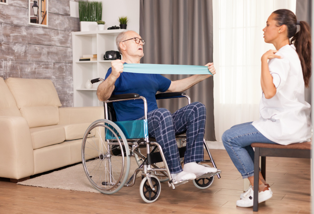
Stroke Rehabilitation in Brampton: Recovery Timeline, Contracture Prevention, Foot Drop, Splinting & Physiotherapy Recovering from a stroke can be one of the most challenging journeys for both patients and their families. While every stroke is unique, one thing remains constant: early rehabilitation and proper management significantly improve recovery and long-term independence. At Movement Rehab Physiotherapy and Manual Therapy Clinic, we provide evidence-based neurological rehabilitation focused on restoring movement, preventing complications, and helping individuals regain confidence in their daily lives. What Is a Stroke? A stroke occurs when the blood supply to part of the brain is interrupted, depriving brain cells of oxygen. This can happen due to: Ischemic stroke – a blockage in a blood vessel (most common). Hemorrhagic stroke – bleeding into the brain. Transient Ischemic Attack (TIA) – often called a “mini-stroke,” which is a warning sign that should never be ignored. Depending on the area of the brain affected, a stroke may cause: Weakness on one side of the body Difficulty walking Loss of balance Speech or swallowing problems Memory and cognitive difficulties Vision changes Muscle stiffness or flaccidity Reduced sensation Fatigue The Stages of Stroke Recovery Recovery does not happen overnight. Most people progress through predictable stages, although the timeline varies for each individual. Stage 1 – Acute Flaccidity (First Days to Weeks) Immediately after a stroke, muscles often become flaccid, meaning they are soft, weak, and unable to contract effectively. During this stage patients commonly experience: Complete weakness or paralysis Poor sitting balance Difficulty standing Shoulder instability Foot drop Inability to move the arm or leg voluntarily This is when proper positioning is most important. Gravity Becomes the Enemy When muscles cannot support the limbs, gravity begins pulling joints into poor positions. Examples include: Shoulder The arm hangs downward because the shoulder muscles cannot support its weight, increasing the risk of: Shoulder subluxation Pain Rotator cuff injury Difficulty regaining movement Wrist and Hand The wrist bends downward while the fingers curl into the palm. Without stretching and positioning, the hand gradually becomes difficult to open. Hip The weak leg often rolls outward while lying in bed, affecting alignment and walking later. Knee The knee may remain bent for prolonged periods, gradually reducing extension. Ankle The ankle naturally falls into a pointed position because gravity and calf muscles overpower the weak muscles that lift the foot. This leads to foot drop. Why Early Positioning Matters Good positioning helps: Protect joints Reduce swelling Prevent pressure injuries Maintain muscle length Improve comfort Reduce pain Prepare muscles for recovery Positioning should be maintained: In bed In a wheelchair While sitting During transfers During standing Even small adjustments made throughout the day can prevent long-term complications. Understanding Contractures One of the biggest challenges after stroke is the development of contractures. A contracture occurs when muscles, tendons, ligaments, and surrounding tissues become permanently shortened due to prolonged immobility. Contractures commonly develop in: Shoulder Elbow Wrist Fingers Hip Knee Ankle Once established, contractures can: Cause pain Reduce movement Limit independence Make dressing and hygiene difficult Interfere with walking Require prolonged rehabilitation Occasionally require surgical treatment The best treatment for contractures is prevention. Preventing Contractures An effective prevention program includes: Daily passive range-of-motion exercises Active exercises whenever possible Stretching Proper positioning Standing programs Splints and orthotics Walking practice Regular physiotherapy Occupational therapy Home exercise programs Caregiver education Consistency is far more important than intensity. Foot Drop After Stroke Foot drop occurs because the muscles that lift the foot become weak or lose coordination. Signs include: Toes dragging while walking Tripping frequently High-stepping gait Difficulty climbing stairs Increased fall risk Without treatment, walking becomes inefficient and unsafe. Treatment may include: Strengthening exercises Gait training Balance training Stretching Functional Electrical Stimulation (FES) Ankle-Foot Orthosis (AFO) Manual therapy Task-specific walking practice Spasticity: When Muscles Become Too Tight As the brain begins recovering, many patients transition from flaccidity to spasticity, where muscles become stiff and overactive. Common patterns include: Upper Limb Shoulder pulled inward Elbow bent Wrist flexed Fingers tightly closed Lower Limb Stiff knee Pointed foot Inward-turning leg Difficulty bending the knee during walking Spasticity can interfere with: Walking Dressing Hygiene Sleeping Hand function Transfers Physiotherapy, stretching, splinting, medications, and, in some cases, botulinum toxin injections can help manage spasticity. Why Splinting Is So Important Splints are used to maintain joints in a functional position while muscles recover. Hand Splints Help: Prevent finger contractures Maintain thumb position Reduce pain Improve hand hygiene Protect joints Preserve future function Wrist Splints Help: Maintain wrist alignment Reduce abnormal flexion Improve positioning Ankle-Foot Orthoses (AFOs) Help: Prevent foot drop Improve walking safety Reduce falls Improve balance Increase walking efficiency Night Splints Help maintain muscle length while sleeping and reduce morning stiffness. Splints should always be individually prescribed and regularly reviewed to ensure proper fit and skin protection. Stroke Recovery Scales Used by Physiotherapists Physiotherapists use standardized outcome measures to monitor recovery and guide treatment. Brunnstrom Stages of Recovery Describes progression from flaccidity to coordinated movement. Fugl-Meyer Assessment Considered one of the gold-standard measures for motor recovery following stroke. Measures: Arm movement Leg movement Coordination Balance Sensation Joint movement Modified Ashworth Scale Measures muscle tone and spasticity. Berg Balance Scale Assesses static and dynamic balance and helps determine fall risk. Timed Up and Go (TUG) Measures mobility, transfers, walking, and balance. 10-Metre Walk Test Measures walking speed and predicts community mobility. Six-Minute Walk Test Measures endurance and walking capacity. Functional Independence Measure (FIM) Evaluates independence with: Walking Dressing Bathing Transfers Toileting Self-care Stroke Impact Scale Assesses the effect of stroke on physical function, communication, memory, emotion, participation, and overall quality of life. Neuroplasticity: The Brain Can Relearn The brain has an incredible ability known as neuroplasticity, allowing healthy brain areas to form new connections and take over some lost functions. Neuroplasticity is enhanced by: Repetitive practice Task-specific exercises Walking training Balance activities Functional arm use